
Dr Manisha Bahl is a breast imaging radiologist and past director of the Breast Imaging Fellowship Programme at the Massachusetts General Hospital and Harvard Medical School in Boston. She is working with researchers from MIT’s Computer Science and Artificial Intelligence Laboratory (CSAIL). Their research focuses on applying artificial intelligence (AI) techniques to critical areas in breast cancer detection, diagnosis, and treatment.

“The power of the AI approach is that AI algorithms can continually improve with exposure to more imaging data and have the potential to discover imaging features and relationships among features that are currently unknown to us.”
Bringing together breast imaging and AI
I am a radiologist specialised in breast imaging at Harvard Medical School and the Massachusetts General Hospital (MGH). I completed my studies at Stanford University, the University of California – San Francisco School of Medicine (UCSF), and the Harvard T.H. Chan School of Public Health, and my residency and fellowship training at Duke University Medical Center.
Most recently, I completed the Professional Certificate in Machine Learning and AI at the Massachusetts Institute of Technology (MIT), in order to enhance the quality and impact of my research utilizing cutting-edge technologies to improve clinical care.
Currently, I am a National Institutes of Health (NIH)-funded investigator with research interests in the application of AI to improve outcomes in women with breast cancer and in the clinical assessment of digital breast tomosynthesis (also known as 3D mammography).
I am an associate editor for Radiology: Artificial Intelligence and the Journal of Breast Imaging and podcast co-host for the journal, Radiology, and I also serve on the Scientific Advisory Committee of the Society of Breast Imaging (SBI).
Saving lives through breast imaging
On a day-to-day basis, I provide clinical care in breast imaging, lead research projects to advance the field of breast imaging, and teach and supervise medical students, residents, and fellows.
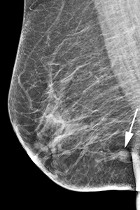
My clinical work involves interpreting breast imaging exams (including mammograms, breast ultrasound, and breast MRI), performing breast biopsies, and consulting with patients and the multidisciplinary breast cancer care team.
Breast cancer diagnoses delayed by COVID
Breast cancer screening was particularly affected by the COVID-19 pandemic. According to an article published in the Journal of Clinical Oncology Clinical Cancer Informatics, screening mammography dropped by 90% at the peak of the pandemic when compared to the same time period last year.
It is estimated that more than 35,000 breast cancer diagnoses could be delayed and that 5,200 more women may die in the United States over the next decade as a result of the screening hiatus, according to a report from the IQVIA Institute for Human Data Science and an article from the journal, Science. Fortunately, many restrictions due to the pandemic have now been lifted where I work in Massachusetts, and many of our patients have resumed breast cancer screening.
How AI can improve clinical efficiency in breast imaging practices
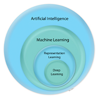 AI is a branch of computer science dedicated to developing computer algorithms that imitate intelligent human behaviour. It encompasses machine learning, in which the computer learns from data without being programmed, and deep learning, in which neural networks are used to extract high-level features from raw data.
In the domain of breast imaging, an AI algorithm can teach itself what a breast cancer looks like by extracting and learning imaging features from provided images of breast cancers. The power of the AI approach is that AI algorithms can continually improve with exposure to more imaging data and have the potential to discover imaging features and relationships among features that are currently unknown to us.
AI algorithms for breast imaging have been shown to improve radiologists’ accuracy in detecting breast cancers on mammography, reduce false-positive exams, quickly identify cancer-free mammograms, decrease interpretation times, accurately assess an individual woman’s risk of breast cancer, and provide early prediction of neoadjuvant chemotherapy response, among many other applications.
The many usages of AI in breast imaging
AI in medicine has rapidly advanced from pilot and feasibility studies to implementation into clinical practice.
In breast imaging, AI systems can be used for automated interpretation, triage, or decision support:
- With regard to automated interpretation, AI systems have been shown to improve radiologists’ accuracy in detecting breast cancers on mammography.
- A triage approach would involve the AI system running in the background to identify cases that need to be evaluated more urgently.
- With regard to decision support, AI systems can be used to accurately assess an individual woman’s risk of breast cancer and therefore inform personalised decisions about screening regimens and prevention strategies. AI systems can also be useful to guide clinical decision-making with regard to surgical excision versus surveillance of certain types of breast lesions.
Eliminating bias in AI algorithms
As an associate editor for Radiology: Artificial Intelligence, I recently had the opportunity to attend the Fourth Annual Conference on AI, Ethics, and Society, organised by the Association for the Advancement of Artificial Intelligence (AAAI) and the Association for Computing Machinery (ACM).
One of the themes addressed in this year’s conference was bias in AI algorithms, which is of particular interest to me as a researcher focused on developing and implementing AI algorithms into clinical practice.
Biases in AI algorithms can arise from various sources, such as incomplete data, unrepresentative data, and historical biases. For example, research has shown that AI-based facial analysis algorithms are more likely to misclassify minorities, especially minority women, likely because of unrepresentative training data used to develop the algorithms.
Detection and mitigation of such biases are critical for us to be able to trust and therefore use AI algorithms in clinical practice and in other settings.
People from different fields must work together
Multidisciplinary collaboration is essential for the successful development and implementation of AI algorithms into clinical practice. The unique skillsets, experiences, and perspectives brought by each member of the multidisciplinary team can lead to solutions to challenging problems in medicine and other domains that are simply not possible without collaboration.
For example, my current work on the use of AI to risk-stratify women with stage 0 breast cancer (an early-stage breast cancer, known as ‘precancer’ or ductal carcinoma in situ [DCIS]) is being done in collaboration with experts in AI, biostatistics, radiology, breast surgery, breast pathology, and implementation science.
Helping women making more informed decisions
In 2019, I was fortunate to receive more than $1.3 million in funding from the National Institutes of Health (NIH) to develop and implement an AI algorithm that accurately risk-stratifies women with ductal carcinoma in situ (DCIS), also known as stage 0 breast cancer.
This work is in progress and is being done in collaboration with a multidisciplinary team that includes experts from MIT (Massachusetts Institute of Technology), Massachusetts General Hospital, and Harvard.
The AI algorithm will combine clinical, imaging, and pathology data, using cutting-edge machine learning and deep learning techniques, and I believe that the use of the AI algorithm in the clinical setting will empower women with DCIS to make more informed decisions with regard to their treatment options.
I hope to continue leading collaborative research efforts to investigate and apply AI techniques to other critical areas in breast cancer detection, diagnosis, and treatment.
https://www.linkedin.com/in/manisha-bahl-a44a92a1/
https://www.massgeneral.org/doctors/19887/Manisha-Bahl